Research
Overview: Self-Assembly, Soft Devices, Active
Matter, Cells and Soft Glasess
|
Our research interests are in soft matter physics and
engineering and their border with cell biology. Our
expertise lies in figuring out how to make careful
mechanical measurements on very small objects, from
colloidal particles and cells all the way down to single
macromolecules. On the one hand, we can use biochemistry to
construct novel and interesting soft-matter experiments and
materials and perhaps ultimately to engineer useful devices.
On the other hand, we can use methods originally developed
for studying soft matter to probe interesting biological
systems, such as the cytoskeleton of eukaryotic cells.
|
Directed
Self-Assembly and Active Micro-devices by Molecular
Recognition
|
The term self-assembly describes the spontaneous
organization of component parts, for example, atoms
arranging themselves into an ordered crystal. Much attention
has been placed on the idea of 'directed self-assembly' to
‘grow’ useful devices from nanoscopic synthetic parts (also
called ‘bottom up’ assembly) rather than current
nanofabrication techniques based on photolithography (‘top
down’ assembly). Ideally, we would like to have a technology
platform that allows us to self-assemble arbitrary,
complicated structures from a large number of different
parts, and to do so rapidly and with a low probability of
forming defective assemblies. In principle, this would seem
to require parts that exclusively stick to each other in a
user specified way. In practice, one of the most successful
approaches is due originally to Ned
Seeman. This approach, called DNA
nanotechnology, uses DNA to stick structural components
together by exploiting the tendency of single stranded DNA
to form a double helix preferentially with strands having a
complementary base sequence. While this method has been used
by a number of researchers to form an ever increasing array
of complicated structures, the details of why some designed
structures form while others do not remain poorly
understood.
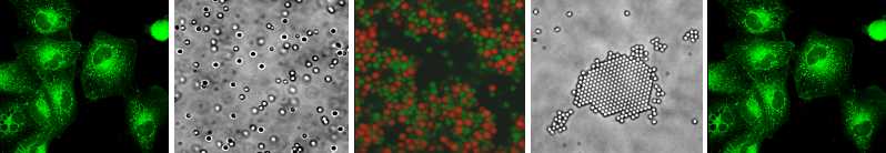
In our lab, we apply DNA nanotechnology to self-assemble
novel structures from micron-sized objects, specifically
micron-sized polymer spheres called colloids. We chose
colloids because the formation of ordered colloidal crystals
is a comparatively well understood and prototypical example
of self-assembly, and colloids are large enough to be imaged
using an optical microscope, making it relatively easy both
to watch the dynamics assembly process as it occurs, and to
see what you made in the end. We were the first group to
successfully form colloidal crystals using interactions due
to DNA (or any molecular recognition system). In the end, we
found issues of colloidal stability, reversible binding,
equilibrium growth and annealing to be critically important.
Along the way we developed a novel method for modifying the
surface of colloids with a polymer brush and functional
groups.
Our current efforts are directed at forming novel two and
three dimensional alloy structures. This exploratory
research receives input from detailed simulations by Talid Sinno’s group.
In our original approach, we prepare a mixture of different
particles, A, B and C, having specific interactions (e.g. A
sticks to B but not to C, etc.) which we describe by an
interaction matrix. By adjusting the number of
species, their size, and the interaction matrix a variety of
different crystal and cluster symmetries can be
self-assembled. In a second newer approach, we are
investigating the role that particle shape plays in defining
the final structure. We currently produce
non-spherical building blocks using a nano-molding method we
call 'crystal templating' that uses a colloidal crystal as a
general-purpose mold for building other shapes.
In a separate project with Daeyeon
Lee and David
Chenoweth, we are using a technique called Layer
by Layer deposition to create thin, flexible films of
DNA-grafted gold nanoparticles. By placing DNA
nano-actuators in these films, we can create films that
reversibly fold themselves into new shapes, driven by DNA
fuel strands. Current efforts are directed at
patterning these new active materials, and building novel
micro-origami structures and simple micro-robotic devices.
|
Binding in
the Single- and Few-Molecule Limit
|
As a spin-off of our self-assembly work, we have performed
direct measurements of the effective pair potential of mean
force prevailing between two colloidal particles bearing
complimentary DNA. This effort required the refinement of an
extended optical tweezer measurement system to achieve
resolution at the few nanometer level. Direct measurements
of the DNA induced interaction were well described by a
semi-analytic model we developed based on polymer physics,
chemical equilibrium and a generalized mass action
principle. While our first measurements were at high DNA
density (up to ~100 molecules could sterically reach between
the colloids), we soon realized that our instrument was
equally well suited to studying binding in the single or few
molecule limit as well. That work confirmed what many have
begun to suspect—that the kinetics of DNA hybridization near
the melting temperature are non-exponential.
|
Cell
Mechanics and Soft Glasses
|
It is increasingly clear that mechanical stress and
deformation play a significant role in the behavior of
eukaryotic cells. It also appears likely that cells are
mechanically sophisticated, having an array of mechanical
senses whose outputs are integrated together and used during
differentiation decisions, tissue remodeling and, of course,
motility. Understanding these aspects of cell mechanics
would seem to be a prerequisite both for understanding
tissue growth and morphology at the cellular scale and
realizing the ultimate potential of tissue engineering.
Despite this potential impact, the mechanical response (e.g.
rheology) of cells and its physical and biochemical
determinants remain largely mysterious. In our previous
work, we applied a variety of different microrheology
techniques to understand cell mechanics and rheology. The
challenge of understanding cell mechanics is a consequence
of their mechanical complexity. In a series of studies
applying different microrheological methods to the same
cells, we demonstrated that cells' different compartments
and discrete structural elements each had their own distinct
mechanical response. In general, different measurement
techniques measure different components, or different
combinations of components, and can do so in a cell type
dependent manner.
In our current research, we use a a unique tool for
quantifying the stress fluctuations within a soft material,
which we call stress fluctuation spectroscopy, developed
with our collaborators Andy Lau and Tom Lubensky. A major
current area of soft-matter research relates to
understanding materials containing active elements that
cause internal deformation, sliding or stress, for example,
molecular motors. While cells are obviously an example of
such materials, how quantities such as motor activity and
intracellular stress relate in general to the mechanical
response and function remain unclear. Given the array of
different motors and stress generation mechanisms in cells,
it seems likely that the observed basal stress and stress
fluctuations will be a superposition of many different
sources, some of which may be intimately related to the
mechanics and others that are not. Current efforts are
directed at understanding the molecular mechanisms and
biophysics of neutrophil spreading with Dan
Hammer and endothelial cell crawling and
chemokinesis with Dan
Reich at Johns Hopkins.
The mechanics of cell structures have distinctive,
viscoelastic mechanical properties that closely resemble
Soft Glassy Materials, a class of non-biological materials
including foams, dense emulsions (like mayonnaise) and
slurries (like ketchup), that are neither completely liquid
nor solid. The physical origin of these
materials distinctive mechanics remains mysterious despite
long study. In a collaboration with Rob
Riggleman, we are building novel computational
models of such materials and relating their rheology to the
nature of the unusual relaxation pathways the system takes
when particles rearrange.
|